
Imagine you are sitting on the sofa, watching a movie, when suddenly your chest tightens. You try to take a deep breath, but it feels like someone has clamped your airway shut. Your heart starts racing. This isn't just "being out of shape." It could be a pulmonary embolism, which is a life-threatening blockage in one or more arteries in the lungs caused by blood clots.
This condition strikes without warning for many people. In fact, sudden shortness of breath is the most common sign, appearing in 85% of cases according to data from the National Institutes of Health (NIH). Because the symptoms can mimic anxiety, asthma, or even a heart attack, diagnosis is often delayed. Understanding what happens inside your body and how doctors identify this emergency can save lives.
What Is a Pulmonary Embolism?
To understand a pulmonary embolism (PE), you first need to look at your legs. Most PEs start as deep vein thrombosis (DVT). This is a blood clot that forms in the deep veins of your lower limbs. About 70% of confirmed PE cases originate from these leg clots. When a piece of that clot breaks loose, it travels through your bloodstream, up to your heart, and then gets stuck in the pulmonary arteries-the vessels responsible for carrying blood from your heart to your lungs for oxygenation.
When these arteries are blocked, blood cannot reach the lung tissue effectively. This disrupts oxygen exchange. Your body screams for more air, leading to dyspnea (shortness of breath). The severity depends on where the clot lands. A small clot in a peripheral artery might cause mild, transient breathlessness. A large clot blocking a main pulmonary artery causes massive PE, a critical vascular emergency where profound shortness of breath occurs even while resting.
The Hallmark Symptom: Sudden Shortness of Breath
If there is one symptom you must recognize, it is abrupt, unexplained difficulty breathing. The NIH reports that 92% of patients with massive PE experience this abruptly. It doesn’t matter if you have been running a marathon or sitting still; the onset is often instantaneous.
However, shortness of breath rarely travels alone. Other symptoms frequently accompany it:
- Chest pain: Occurs in 74% of cases. Unlike the crushing pressure of a heart attack, this pain is often pleuritic-meaning it is sharp and worsens when you take a deep breath, cough, or bend over. The Mayo Clinic notes that 68% of patients describe it as localized and sharp.
- Coughing: Happens in 53% of cases. Sometimes, you may cough up small amounts of blood (hemoptysis), which occurs in about 23% of patients.
- Rapid heart rate: Tachycardia affects 30% of patients, with heart rates exceeding 100 beats per minute.
- Fainting: Syncope occurs in 14% of cases, usually signaling a significant drop in blood pressure due to the heart struggling to pump against the blockage.
- Leg swelling: If the source is a DVT, you might notice swelling, warmth, or pain in one leg (44% of cases).
A crucial diagnostic clue is "unexplained hypoxemia." This means your blood oxygen levels are low, but a standard chest X-ray looks normal. If you have low oxygen and a clear chest X-ray, doctors should immediately suspect PE.
Who Is at Risk?
Pulmonary embolism does not discriminate, but certain factors significantly raise your odds. The American Lung Association estimates that PE results in approximately 100,000 deaths annually in the United States, with an incidence of 60-70 cases per 100,000 people.
Your risk skyrockets if you have:
- Cancer: Patients with cancer face a 4.7-fold increased risk of PE compared to the general population, according to the European Society of Cardiology.
- Recent surgery or trauma: Especially orthopedic surgeries like hip or knee replacements.
- Immobility: Long-haul flights, hospital stays, or bed rest allow blood to pool in the legs.
- History of clots: Dr. Gregory Piazza from Harvard Medical School notes that 33% of patients with prior DVT or PE will experience a recurrence within 10 years.
- Hormonal factors: Birth control pills or hormone replacement therapy can increase clotting tendency.
How Doctors Diagnose Pulmonary Embolism
Diagnosing PE is a race against time. Because symptoms overlap with so many other conditions, doctors use a structured, multi-step approach validated by the American Thoracic Society.
Step 1: Clinical Probability Scores
Before ordering expensive scans, clinicians assess your likelihood of having a PE using scoring systems like the Wells Criteria or the Geneva Score. These tools ask questions about your symptoms, vital signs, and risk factors. They help categorize you as low, moderate, or high probability. The NIH reports that when applied by trained clinicians, these tools correctly identify low-risk patients in 89% of cases.
Step 2: The D-Dimer Blood Test
If your clinical probability is low or moderate, you will likely get a D-dimer test. D-dimer is a protein fragment produced when a blood clot dissolves. A negative result (less than 500 ng/mL) rules out PE with 97% sensitivity in low-risk patients. However, this test has a major flaw: it lacks specificity. It can be elevated due to infection, inflammation, pregnancy, or simply being over age 50. For patients over 50, the specificity drops from 94% to 54%, meaning a positive result often requires further imaging regardless of the score.
Step 3: Imaging Confirmation
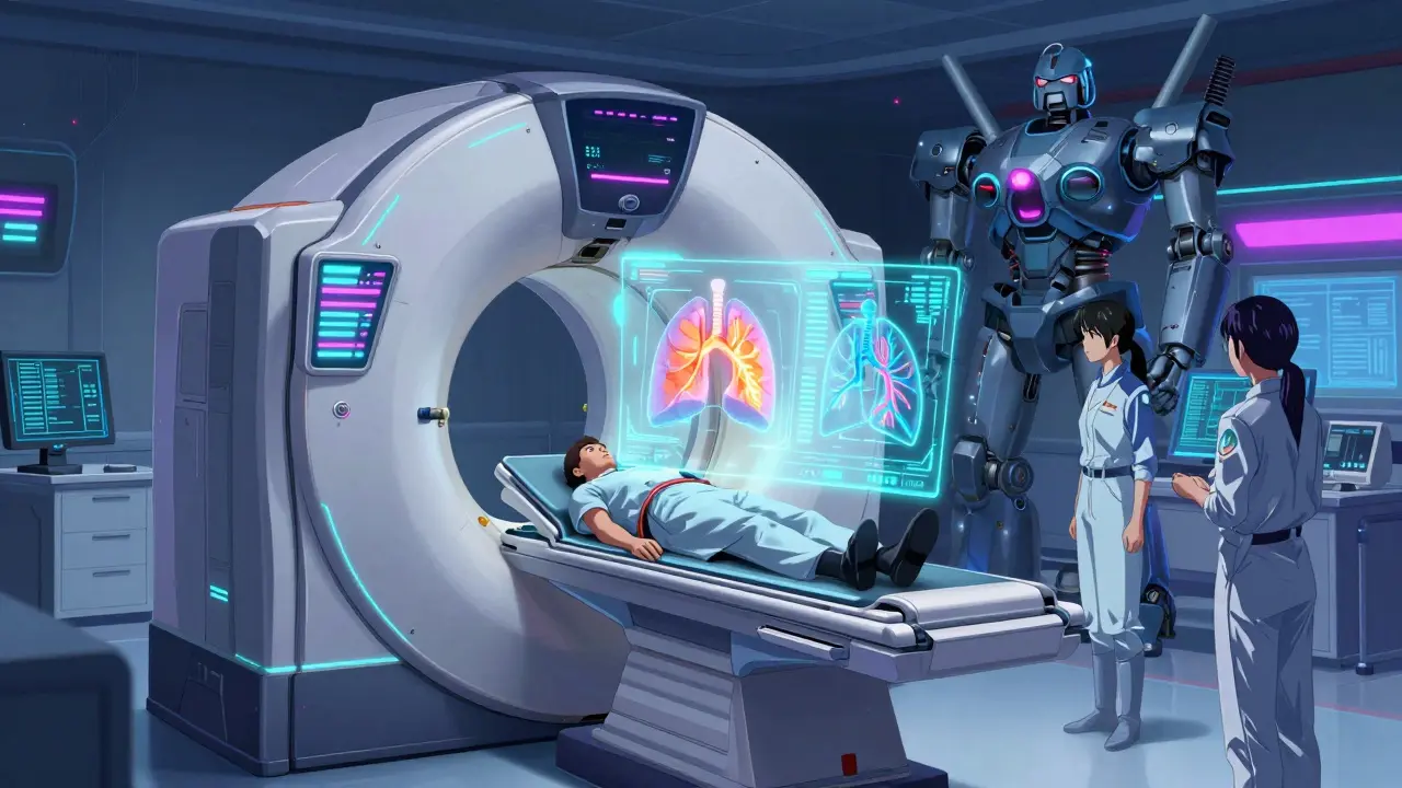
If the D-dimer is positive or your clinical risk is high, imaging is mandatory. The gold standard is Computed Tomography Pulmonary Angiography (CTPA).
| Test | Sensitivity | Specificity | Best Used For |
|---|---|---|---|
| CTPA (CT Scan) | 95% | 96% | Gold standard; most patients |
| V/Q Scan | 85% | 95% | Pregnant women or those with contrast allergies |
| Compression Ultrasound | >90% | 95% | Detecting DVT in legs if lung imaging is inconclusive |
| Echocardiogram | 84% | N/A | Bedside assessment for unstable patients |
During a CTPA, you receive iodinated contrast dye through an IV. The scanner takes detailed images of your lung arteries. It detects PE in 92% of cases with high precision. If you cannot have contrast dye (due to kidney issues or allergy), doctors may order a Ventilation/Perfusion (V/Q) scan, which compares airflow and blood flow in your lungs.
For patients who are hemodynamically unstable (e.g., fainting, very low blood pressure), doctors skip the CT scanner and go straight to a bedside transthoracic echocardiogram. This ultrasound of the heart checks for right ventricular strain, a sign that the heart is struggling against the blockage.
Why Diagnosis Is Often Delayed
Despite advanced technology, misdiagnosis remains a serious problem. A survey by Healthdirect Australia found that 68% of PE patients visited healthcare providers an average of 2.3 times before getting the correct diagnosis. Many were initially told they had pneumonia, asthma, or anxiety.
One patient, 'SarahK_42', shared her story on the American Lung Association forum: she experienced sudden shortness of breath climbing stairs for three weeks. Doctors dismissed it as anxiety until she nearly fainted. Another user on Reddit described sudden breathlessness while watching TV, only to be taken seriously after a fainting episode.
These delays happen because PE is a "great mimicker." Its symptoms are nonspecific. Furthermore, rural areas often lack access to V/Q scanning or rapid CTPA capabilities. The Bupa UK guidelines note that specialized nuclear medicine facilities are available at only 78% of major hospitals, creating disparities in care speed.

New Advances in Detection
Medicine is evolving to catch these clots faster. The American College of Chest Physicians introduced age-adjusted D-dimer thresholds in 2023. Instead of a flat cutoff, the threshold increases by 10 ng/mL for every year over age 50. This simple change reduced unnecessary imaging by 36.4% in a trial of 14,000 patients, sparing healthy older adults from radiation exposure.
Artificial intelligence is also stepping in. Algorithms like PE-Flow are now assisting radiologists in interpreting CTPA scans, demonstrating 93.7% sensitivity in recent trials. Additionally, multidisciplinary Pulmonary Embolism Response Teams (PERTs) are becoming standard in major hospitals. These teams coordinate care instantly, reducing time-to-treatment by 3.2 days and lowering mortality in massive PE cases.
What Should You Do?
If you experience sudden, unexplained shortness of breath, especially if accompanied by chest pain, rapid heartbeat, or leg swelling, do not wait. Do not assume it is stress or indigestion. Seek emergency medical attention immediately. Mention your risk factors clearly to the triage nurse. Early diagnosis leads to early treatment with anticoagulants (blood thinners), which prevents the clot from growing and stops new ones from forming.
Can a pulmonary embolism happen without leg pain?
Yes. While 70% of PEs originate from deep vein thrombosis (DVT) in the legs, many patients do not experience noticeable leg pain or swelling before the clot travels to the lungs. The absence of leg symptoms does not rule out PE, especially if you have sudden shortness of breath.
Is a D-dimer test enough to diagnose PE?
No. A negative D-dimer test can rule out PE in low-risk patients, but a positive result is not definitive. Because D-dimer levels can rise due to age, infection, or inflammation, a positive result always requires confirmation with imaging like a CTPA or V/Q scan.
How long does it take to get diagnosed with PE?
In ideal settings with established protocols, diagnosis can occur within 45 minutes via rapid CTPA pathways. However, real-world data shows many patients see multiple doctors over days or weeks due to misdiagnosis as asthma or anxiety. Immediate presentation to an ER with clear symptoms speeds up the process.
What is the difference between a heart attack and a pulmonary embolism?
Both cause chest pain and shortness of breath. Heart attack pain is often described as crushing pressure radiating to the arm or jaw. PE pain is typically sharp and worsens when you take a deep breath (pleuritic). Only medical testing (ECG, troponin levels, and CT scans) can definitively distinguish between them.
Are there natural ways to prevent blood clots?
While no natural remedy replaces medical prevention for high-risk individuals, lifestyle changes help. Stay hydrated, move regularly during long trips (walk every hour), avoid prolonged immobility, and maintain a healthy weight. If you have a history of clots, follow your doctor's prescription for anticoagulants strictly.
