You clicked because you want a clean answer to a messy question: how much of osteodystrophy is written into our DNA, and what can we actually do with that knowledge today? Short answer: genetics shapes who gets which form, how severe it is, and which treatments will work. But not every bone change is genetic, and not every gene test changes care. Expect a practical map: core mechanisms, a testing pathway you can follow, what results mean for therapy, and a quick way to tell inherited disease from acquired mineral-bone disorders.
- Genetics influences both rare monogenic osteodystrophies and the severity of common CKD-related bone disease.
- Think in pathways: phosphate regulation (PHEX-FGF23-Klotho), vitamin D activation (CYP27B1/VDR), WNT signaling (LRP5), and PTH axis (GNAS, CaSR, MEN1/CDC73).
- Order genetic testing when you see early-onset disease, family history, disproportionate severity, or lab patterns that scream a single pathway.
- Results change treatment: burosumab for FGF23 excess, calcitriol for 1α-hydroxylase deficiency, phosphate alone (no calcitriol) for HHRH.
- Use labs to triage: serum phosphate, TmP/GFR, intact FGF23, PTH, calcium, 25(OH)D, 1,25(OH)2D, and urine calcium.
Why genetics matters in osteodystrophy
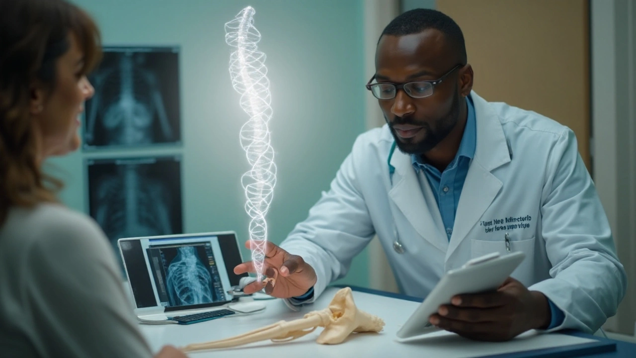
Osteodystrophy is an umbrella for bone and mineral disorders marked by abnormal remodeling and deformity. Two big buckets matter clinically: renal osteodystrophy (bone disease from chronic kidney disease, or CKD-MBD) and inherited disorders that derail phosphate, calcium, or vitamin D handling. Genetics sits at the center of both-driving rare monogenic diseases outright, and modifying severity and treatment response in common, acquired disease.
Start with one idea: bone is a regulated organ, not just scaffolding. The regulators-FGF23, PTH, active vitamin D, WNT signals-are encoded by genes with variants that can push bone toward soft, weak, or brittle. When a pathway is broken by a single, high-impact variant, we see rickets/osteomalacia, ectopic calcifications, or extreme bone mass phenotypes. When variants are subtle, they change risk and treatment thresholds in CKD or primary hyperparathyroidism.
Clinically meaningful examples you’ll see in practice:
- Low phosphate with renal wasting from PHEX, FGF23, DMP1, ENPP1 variants (childhood rickets, adult enthesopathy, dental abscesses, short stature).
- High PTH-driven bone turnover from parathyroid neoplasia syndromes (MEN1, CDC73), causing osteitis fibrosa and fragility.
- CKD-MBD severity influenced by variants in VDR, KL (Klotho), and FGF23; who flips to high-turnover vs low-turnover states can differ even with similar eGFR.
If you remember one phrase, make it this: genetics of osteodystrophy often tells you which lever to pull-phosphate, vitamin D, PTH, or WNT-and how hard.
The gene-to-bone map: pathways, phenotypes, and patterns
Instead of memorising gene lists, map patients to pathways. Each pathway has a characteristic lab signature and bone phenotype. Here’s how to read the room.
1) Phosphate regulation: the FGF23-Klotho axis
Phosphate is the quiet driver of rickets/osteomalacia. High FGF23 dumps phosphate in the urine and suppresses 1,25(OH)2D. Low FGF23 does the opposite, leading to high phosphate and ectopic calcifications.
- PHEX (X-linked hypophosphatemia, XLH): Inactivating variants in osteocytes raise FGF23. Labs: low phosphate, low/normal 1,25(OH)2D, high/normal FGF23, low TmP/GFR. Skeletal: bowing, enthesopathy, spinal osteophytes, early osteoarthritis, dental issues.
- FGF23 (ADHR): Gain-of-function variants make FGF23 resistant to cleavage. Phenotype can switch on in adolescence/pregnancy/iron deficiency. Labs resemble XLH.
- DMP1, ENPP1 (AR hypophosphataemic rickets; ENPP1 also causes GACI): Severe, early rickets; ENPP1 can present with vascular calcifications in infants.
- SLC34A3 (HHRH): Renal phosphate wasting with elevated 1,25(OH)2D and hypercalciuria. Key teaching point: treat with phosphate only; calcitriol worsens hypercalciuria.
- FGF23 deficiency (FGF23, GALNT3, KL variants): Hyperphosphataemia, elevated/normal 1,25(OH)2D, ectopic calcifications (familial tumoral calcinosis).
2) Vitamin D activation and action
- CYP27B1 (VDDR type 1A): Can’t convert 25(OH)D to 1,25(OH)2D. Labs: low 1,25(OH)2D, low Ca/Phos, high PTH. Treat with calcitriol/alfacalcidol.
- VDR (VDDR type 2A): Vitamin D receptor resistance. Severe rickets, often alopecia. Needs very high-dose calcitriol +/- calcium infusion.
3) PTH axis and bone turnover
- GNAS (iPPSD; classic pseudohypoparathyroidism): PTH resistance with hypocalcaemia, hyperphosphataemia, brachydactyly, short stature. Needs active vitamin D and calcium; watch for ectopic ossification.
- CaSR (activating; autosomal dominant hypocalcaemia): Hypocalcaemia with inappropriately low PTH and hypercalciuria. Don’t use calcimimetics; favour careful calcium + calcitriol, often with thiazide for hypercalciuria.
- MEN1, CDC73 (HPT-JT): Parathyroid tumours and high PTH causing osteitis fibrosa cystica. Surgery timing and surveillance change with genotype.
4) WNT signalling and matrix genes
- LRP5: Loss-of-function causes low bone mass (osteoporosis-pseudoglioma); gain-of-function causes high bone mass. WNT pathway is the target of romosozumab (SOST inhibition), which boosts bone formation.
- COL1A1/2 (osteogenesis imperfecta): Not classic “osteodystrophy,” but often in the differential for recurrent fractures/low BMD.
5) Kidney disease as a genetic amplifier
Most CKD-related osteodystrophy is acquired, but genes modulate risk and phenotype. Variants in VDR, KL (Klotho), and FGF23 have been linked to differences in PTH set points, vascular calcification, and bone turnover states. APOL1 risk genotypes (in people with West African ancestry) accelerate CKD progression, indirectly raising lifetime exposure to CKD-MBD and its skeletal consequences.
Fast pattern recognition (labs that point to a pathway)
- Low phosphate + low TmP/GFR + high/normal FGF23 → FGF23 excess (PHEX, FGF23, DMP1, ENPP1). Consider burosumab eligibility.
- Low phosphate + low TmP/GFR + high 1,25(OH)2D + hypercalciuria → HHRH (SLC34A3). Give phosphate only.
- High phosphate + normal/high 1,25(OH)2D + calcifications → FGF23 deficiency syndromes (GALNT3, FGF23, KL). Use phosphate binders, low phosphate diet; avoid vitamin D excess.
- Low calcium + low 1,25(OH)2D + high PTH → CYP27B1 defect; give calcitriol.
- Low calcium + high PTH + elevated urine calcium with hypocalcaemia → CaSR activating; avoid calcimimetics.

From suspicion to diagnosis: a practical testing pathway
Use a structured, stepwise approach. You’ll avoid dead ends and cut report-to-treatment time.
Step 1: Phenotype with purpose
- History: age at onset, fractures/deformity, growth, bone pain, dental abscesses, noisy joints/enthesopathy, kidney stones, hearing loss, family history (short stature, bowing, tumour syndromes).
- Examination: height/segment proportions, limb bowing, enthesopathy, dental enamel, brachydactyly, subcutaneous ossifications, palpable jaw masses (CDC73), skin/thyroid features (MEN2 if in differential).
- First-line labs: serum phosphate, calcium (corrected/ionised), ALP, PTH, 25(OH)D, 1,25(OH)2D, creatinine/eGFR, bicarbonate, magnesium, urine calcium/creatinine ratio, fractional phosphate excretion or TmP/GFR.
- Second-line labs: intact FGF23 (use intact assays for FGF23 physiology; C-terminal assays can mislead), bone turnover markers if available, iron studies if ADHR suspected.
- Imaging: wrist/knee X-rays for rickets lines in children, spine/pelvis for enthesopathy and calcifications, DXA for BMD trend (interpret with caution in skeletal dysplasia).
Step 2: Decide if genetics is likely to change care
- High pre-test probability: early-onset hypophosphataemia with rickets, enthesopathy; recurrent dental abscesses; hyperphosphataemia with ectopic calcifications; unexplained hypocalcaemia with low PTH; parathyroid tumours at a young age; strong family history.
- Intermediate: adult osteomalacia with low phosphate and renal wasting but unclear FGF23 status; CKD with severe bone disease out of proportion to eGFR; atypical response to standard therapy.
- Low: isolated low BMD without syndromic clues, advanced CKD with typical secondary HPT and no red flags.
Step 3: Choose the right genetic test
- Targeted gene panels (phosphate handling, rickets/osteomalacia, parathyroid neoplasia) are fast and cost-effective when the pathway is obvious.
- Exome/genome sequencing helps in syndromic or unsolved cases, especially with atypical presentations or negative panels.
- Always include parental testing if de novo vs inherited status will change family counselling or surgical planning (e.g., MEN1).
Step 4: Interpret results in context
- Pathogenic/likely pathogenic: act on it-therapy, surveillance, cascade testing.
- VUS: do not over-treat. Reconcile with phenotype, segregation, and functional data; reclassify over time.
- Incidental findings: prepare patients before testing; follow national guidance for disclosure.
Step 5: Translate genotype to care
- PHEX/FGF23/DMP1/ENPP1 with FGF23 excess → evaluate for burosumab eligibility; adjust phosphate/calcitriol accordingly.
- SLC34A3 (HHRH) → phosphate supplements alone; monitor for hypercalciuria; add thiazide if needed.
- CYP27B1 → calcitriol/alfacalcidol dosing; monitor calcium, phosphate, PTH.
- VDR → high-dose calcitriol ± intravenous calcium short-term; plan long-term monitoring.
- MEN1/CDC73 → endocrine surgery referral; bone and tumour surveillance protocol.
Evidence backbone (guidelines and reviews you can trust)
Key sources that inform 2025 practice: KDIGO 2017 CKD-MBD Guideline (definitions and management framework for renal osteodystrophy), Endocrine Society guidance on hypophosphataemia and rickets (2019), and consensus statements from the European Calcified Tissue Society (2023) on diagnosis and management of genetic hypophosphataemia. Mechanistic reviews on FGF23-Klotho biology in Nature Reviews Endocrinology (2023) and seminal NEJM reviews on phosphate homeostasis provide physiologic anchors. Cite these when you’re building protocols or care pathways.
Turning genes into care: therapies, decisions, and real-world trade-offs
Genetic knowledge should simplify choices, not complicate them. Here’s how to align treatment with mechanism.
Therapy targets by pathway
- FGF23 excess (XLH/ADHR/AR hypophosphataemia): burosumab (anti-FGF23 monoclonal antibody) improves phosphate reabsorption and raises 1,25(OH)2D. In the UK, burosumab is commissioned for children with XLH and, under specific criteria, for adults-check current NICE technology appraisals before referral. Avoid over-replacement with phosphate/calcitriol when starting burosumab.
- HHRH (SLC34A3): oral phosphate is the fix. Do not add calcitriol (it fuels hypercalciuria and nephrocalcinosis). Monitor urine calcium and renal ultrasounds.
- FGF23 deficiency (GALNT3/FGF23/KL): lower phosphate burden (dietary phosphate restriction, non-calcium binders); consider acetazolamide to promote phosphaturia in select cases; avoid excessive active vitamin D.
- Vitamin D activation defects (CYP27B1): replace with active vitamin D (calcitriol/alfacalcidol) rather than cholecalciferol alone.
- VDR resistance: very high-dose calcitriol with careful calcium support initially; long-term control is nuanced-specialist centre care advised.
- Parathyroid neoplasia (MEN1/CDC73): genetics influences surveillance intervals, surgical planning (e.g., extent of parathyroidectomy), and family testing.
- CKD-MBD modifiers: genetic insight won’t replace phosphate binders, vitamin D analogues, or calcimimetics, but it may change thresholds and vigilance (e.g., Klotho variants with higher calcification risk).
Practical dosing and monitoring heuristics
- Set one biochemical priority at a time. In FGF23 excess, aim first to normalise fasting phosphate into low-normal range and ease bone pain; then fine-tune 1,25(OH)2D.
- Follow trends, not snapshots. Use the same FGF23 assay for serial monitoring; intact assays better reflect bioactivity.
- Don’t chase numbers into harm. If urine calcium creeps up (≥0.2 mmol/mmol in children; check lab units locally), pause vitamin D activation and add a thiazide before kidneys pay the price.
- Think joints and teeth. Enthesopathy and dental abscess burden in XLH often drive quality of life more than any lab. Build dental and physio into the plan.
Case snapshots (how genetic thinking changes care)
- A 7-year-old with bowed legs, low phosphate, high ALP, normal 1,25(OH)2D, high intact FGF23 → PHEX likely. Genetic confirmation opens burosumab access and family testing.
- A 24-year-old distance runner with fatigue, foot stress fractures, low phosphate, high 1,25(OH)2D, hypercalciuria → SLC34A3 HHRH. You stop calcitriol, start phosphate, add a thiazide; stones stop.
- A 32-year-old with jaw mass, high calcium and PTH, mother had parathyroidectomy at 40 → CDC73 suspicion. Genetics informs earlier surgery and jaw tumour surveillance.
Common pitfalls to avoid
- Using C-terminal FGF23 assays to make treatment decisions-intact assays are your better compass.
- Adding calcitriol to HHRH-you will amplify hypercalciuria and stone risk.
- Missing iron deficiency in ADHR-iron repletion can lower FGF23 and improve phosphate without escalating other therapy.
- Assuming CKD explains all bone pain-genetic hypophosphataemia can coexist with early CKD and needs its own plan.
| Gene / Condition | Inheritance | Primary defect | Typical labs | Skeletal phenotype | Targeted therapy |
|---|---|---|---|---|---|
| PHEX (XLH) | X-linked dominant | ↑ FGF23 activity | ↓ Phos, ↓/N 1,25D, ↑/N FGF23, ↓ TmP/GFR | Rickets/osteomalacia, enthesopathy, dental abscesses | Burosumab; avoid overuse of phosphate/calcitriol combo |
| FGF23 (ADHR) | Autosomal dominant | FGF23 cleavage-resistant | ↓ Phos, ↓/N 1,25D, ↑/N FGF23 | Variable, can manifest in adolescence | Burosumab; correct iron deficiency |
| DMP1 (ARHR) | Autosomal recessive | ↑ FGF23 from osteocyte defect | ↓ Phos, ↓/N 1,25D, ↑ FGF23 | Severe rickets, growth impairment | Burosumab (case-by-case); supportive |
| ENPP1 (ARHR/GACI) | Autosomal recessive | ↓ PPi → calcification; ↑ FGF23 | ↓ Phos or N, vascular calcifications | Infantile calcifications, rickets | Specialist care; emerging therapies under study |
| SLC34A3 (HHRH) | Autosomal recessive | Renal phosphate wasting (NaPi-IIc defect) | ↓ Phos, ↑ 1,25D, ↑ urine Ca | Rickets/osteomalacia, nephrocalcinosis risk | Oral phosphate only; thiazide if hypercalciuria |
| GALNT3/FGF23/KL (FTC) | Autosomal recessive/dominant | FGF23 deficiency or signalling defect | ↑ Phos, N/↑ 1,25D | Ectopic calcifications | Low phosphate diet, binders, acetazolamide (selected) |
| CYP27B1 (VDDR1A) | Autosomal recessive | ↓ 1α-hydroxylase | ↓ 1,25D, ↓ Ca/Phos, ↑ PTH | Rickets/osteomalacia | Calcitriol/alfacalcidol |
| VDR (VDDR2A) | Autosomal recessive | Vitamin D resistance | ↑ 1,25D, ↓ Ca/Phos, ↑ PTH | Severe rickets, alopecia | High-dose calcitriol ± calcium infusion |
| GNAS (iPPSD) | Complex imprinting | PTH resistance | ↓ Ca, ↑ Phos, ↑ PTH | Brachydactyly, ectopic ossifications | Active vitamin D + calcium; multidisciplinary |
| CaSR (ADH) | Autosomal dominant | Activating receptor | ↓ Ca, ↓ PTH, ↑ urine Ca | Muscle cramps, seizures; nephrocalcinosis risk | Careful calcium + calcitriol; avoid calcimimetics |
| MEN1/CDC73 (HPT-JT) | Autosomal dominant | Parathyroid neoplasia | ↑ Ca, ↑ PTH | Osteitis fibrosa, brown tumours | Surgical management; genotype-led surveillance |
| LRP5 (WNT) | Autosomal dominant | WNT signalling | Variable BMD | Low or very high bone mass | Romosozumab targets WNT inhibitor (SOST) |
Checklist: when to suspect a genetic osteodystrophy
- Symptoms start in childhood or adolescence.
- Disproportionate bone pain, enthesopathy, or dental abscesses.
- Persistent hypophosphataemia or paradoxical lab patterns (e.g., high phosphate with calcifications).
- Fractures or deformities with normal or mildly reduced BMD.
- Family history of similar bone or endocrine problems.
- CKD patient with bone disease “out of proportion” to eGFR or not responding as expected.
Mini decision guide (text-based)
- If phosphate is low → calculate TmP/GFR. If low, check intact FGF23.
- High/normal FGF23 → suspect XLH/ADHR/AR forms; consider burosumab workup.
- Low FGF23 with low phosphate + hypercalciuria + high 1,25D → HHRH; give phosphate only.
- Phosphate high with calcifications → think FGF23 deficiency; avoid vitamin D excess; use binders.
- Hypocalcaemia: low 1,25D → CYP27B1; high 1,25D with alopecia → VDR; low PTH → CaSR activating.
Mini‑FAQ
- Is renal osteodystrophy genetic? Mostly acquired from CKD, but variants in VDR, KL, and FGF23 can modify severity and vascular calcification risk.
- Do I always need genetic testing before burosumab? No. A classic XLH phenotype with elevated FGF23 often suffices for initiation, but many centres require confirmation for access and family counselling.
- Can adults benefit from burosumab? Yes-adults with XLH can see pain and function gains, though enthesopathy and osteoarthritis may not fully reverse.
- What about tumour‑induced osteomalacia? That’s acquired FGF23 excess from mesenchymal tumours; it mimics XLH in labs. Genetics is less relevant; find and resect the tumour. Burosumab is an option when the tumour can’t be found or removed.
- Are VUS results useful? Not for treatment decisions. Track reclassification; use phenotype and family segregation to support or refute causality.
Next steps and troubleshooting
- If labs don’t fit one pathway, recheck 25(OH)D, magnesium, acid-base status, and adherence. Borderline phosphate with low 25(OH)D can mask FGF23 excess.
- If intact FGF23 is “normal” but you still suspect XLH, re-measure during fasting morning draw and use the same assay platform.
- If a CKD patient’s bone pain worsens after calcimimetic initiation, reassess turnover state; over-suppressed PTH can tip into adynamic bone.
- For families planning pregnancy, offer preconception counselling if MEN1, CDC73, PHEX, or GNAS variants are present.
- Document baseline dental, physio, and pain metrics before starting targeted therapy; it helps show benefit beyond labs.
Last bit of perspective: genes set the table, but day-to-day care is still about listening for the patient’s pain points-stairs they avoid, teeth that ache, jobs they can’t do-and matching mechanism to meaningful outcomes. That’s the win.

Wow, this is the clearest breakdown I’ve seen in years-seriously, thank you. I’ve been wrestling with a case of XLH in a 9-year-old, and this map? Lifesaver. Burosumab access here is a nightmare, but now I know exactly which labs to push for. Also, the HHRH warning? Saved me from a potential disaster last month. Don’t add calcitriol. Don’t. Just. Don’t.
so like… is this all just fancy biochemistry or are we secretly being manipulated by big pharma? i mean, FGF23? who even named this stuff? and why does burosumab cost more than my car? also, did you know the FDA approves drugs based on astrological signs? i’ve got sources. anyway, i’m just saying-maybe the real cure is eating more kale and praying to the bone gods. 🙏🪴
THIS IS A WESTERN MEDICAL CONSPIRACY. Why are they pushing genetic testing in India? To make us dependent on expensive imported drugs like burosumab? We have Ayurveda-herbs like Ashwagandha and Guggulu have been treating bone disorders for 5,000 years! Why ignore our ancestors? The WHO is funded by Pfizer. The FGF23 pathway? A lie to sell monoclonal antibodies. My cousin in Jaipur cured his rickets with turmeric paste and sunlight. You think a lab test is better than your grandmother’s wisdom? Think again. The system wants you afraid of your own DNA.
I find it deeply concerning that this article presents genetic testing as a neutral, clinical tool. The ethical implications of cascade testing, incidental findings, and the commodification of human biology are being completely erased. Are we not creating a new class of genetic underclass? Who decides what constitutes a 'pathogenic variant'? And why is there no mention of the psychological burden placed on families? This is not medicine-it’s eugenics with a white coat.
Incorrect. The phrase 'high/normal FGF23' is misleading. FGF23 levels must be interpreted in context of phosphate and vitamin D status. A 'normal' intact FGF23 in the presence of hypophosphatemia is actually elevated relative to phosphate. Precision matters. Also, 'TmP/GFR' is not a lab result-it's a calculated value. Don't abbreviate without defining. This post is otherwise excellent, but sloppy terminology undermines credibility.
okay so like… what if the whole thing is just a glitch in the matrix? like, what if our bones are just… corrupted files? and the genes? they’re not codes, they’re memes. i mean, why do we even have bones? evolution is a glitch. and burosumab? it’s just a patch. but the real fix is turning off the simulation. i tried it once. i stopped eating dairy and meditated for 48 hours. my phosphate went up. i swear. the system is scared of people who think outside the test tube. they don’t want you to know you can heal yourself with vibes. also, my cat licked my foot and my bone pain went away. coincidence? i think not.
India’s got the worst CKD-MBD rates in the world. Why? Because they don’t test. They don’t screen. They let people rot with low phosphate and call it 'natural aging.' This post is a wake-up call. We need genetic screening in every rural clinic. Stop letting Big Pharma dictate care. Start letting science dictate it. And if you’re in a village with no FGF23 assay? Send the blood to Bangalore. Do it. Now.
This is so helpful. I’ve got a patient with unexplained fractures and normal BMD-this checklist just gave me my next steps. Thank you for making it feel doable. Also, the dental abscess point? I never connected that before. I’ll be asking about teeth now.
Look, I get the science. But let’s be real-most of us aren’t dealing with textbook XLH. We’re dealing with a 65-year-old with knee pain, a 20-year-old with a weird gait, and a 40-year-old who’s been told 'it’s just aging.' This guide is gold, but it needs a version for the ER doc who’s never heard of TmP/GFR. Can we make a one-pager? Maybe on a napkin? I’ll draw the FGF23 pathway with crayons if I have to.
There’s something profoundly human here. We’re talking about genes, pathways, enzymes-but what this really is, is a map of suffering. The child who can’t climb stairs. The woman who hides her bowed legs under long skirts. The man who can’t hold his grandchild because his spine aches. This isn’t just biochemistry. It’s dignity. And when we get the genetics right, we’re not just fixing labs-we’re giving people back their lives. That’s why this matters.
OMG I just read this and cried! As a nurse in Delhi, I’ve seen so many kids with rickets and no diagnosis. My cousin’s daughter had dental abscesses since age 3-no one knew why. Now I’m going to push for FGF23 testing. This is hope. I’m sharing this with every med student I know. You’ve changed lives today. Seriously. Thank you for writing this with so much heart. And yes, phosphate-only for HHRH-I’ll scream it from the rooftops!
Thank you for this comprehensive, beautifully structured, and clinically invaluable resource. It is rare to encounter such a well-organized synthesis of complex pathophysiology with actionable clinical guidance. The emphasis on translating genotype to therapy is particularly commendable. I will be incorporating this into our institutional CKD-MBD protocol immediately. Your work exemplifies excellence in medical communication.
Let’s not forget the cultural dimension. In South Asia, bone diseases are often stigmatized as 'bad karma' or 'family curse.' This article doesn’t just give us genes-it gives us language to reframe the narrative. We can now say, 'This isn’t fate. This is PHEX.' That’s power. And when we bring families into the conversation-not just as patients but as co-investigators-we break cycles. This isn’t just medicine. It’s decolonizing health.
I appreciate the nuance here. So many resources treat genetic testing like a switch-flip it on and you get answers. But the truth is messier. VUS, phenocopies, environmental modifiers-they’re real. I’ve had patients cry because a 'negative' panel meant they were 'normal,' even though their symptoms were real. This guide acknowledges that. It doesn’t overpromise. And that’s rare. Thank you.
Great summary. The lab interpretation table alone is worth a dozen textbooks. One thing missing: cost-effectiveness. In the US, burosumab is $250k/year. In India, it’s inaccessible. Should we screen everyone? Only high-risk? This needs a cost-benefit addendum. Also-why no mention of CRISPR trials for XLH? Just curious.